
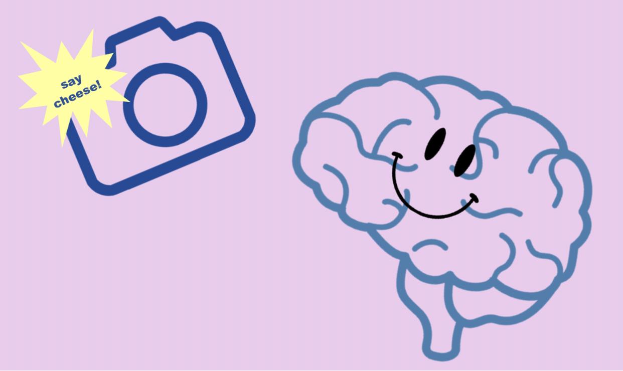
Pictures are fun and easy to take on a phone or camera. But how could you take a picture of your brain? Brain imaging, also called neuroimaging, is very important to doctors and scientists. Pictures of the brain can help doctors detect problems in the brain and make sure it is healthy. But if they can’t use a regular camera, how do they take these images?
There are many different neuroimaging techniques that doctors and researchers can use to image the brain. One of these is Computed topography (CT) scans. If you have ever broken a bone, you have probably gotten an X-ray. X-rays produce black and white images of your bones. The doctors can see if there is a break in the bone. CT scans use X-rays too. They help to show the structure of the brain, but don’t help describe the function of different parts of the brain. Other imaging techniques can do this.
Another type of imaging is a PET scan. No, it’s not like the pets you might have at home. PET stands for Positron Emission Topography. According to Lumen Learning, PET scans measure the level of a sugar called glucose to show where brain activity is occurring. Just like you need to eat a yummy snack to have energy, your brain cells need fuel. This fuel is glucose. When certain areas of the brain are working hard, they are using this sugar more than other areas. Detectors can pick up these general areas, but they cannot pick up specific spots. These detectors not only take images, but they can also take videos of the brain. That being said, they are expensive cameras.
An EEG is also an important imaging tool. EEG stands for electroencephalography. That’s a big word! What an EEG is basically doing is measuring the electrical activities of the brain, according to PsychCentral. It might sound weird that your brain has electrical activity since you use electricity every day for power, but it’s true! EEGs can help doctors diagnose medical problems like seizures. They are often used in research.
One last type of neuroimaging is an MRI. MRI stands for Magnetic Resonance Imaging. MRIs are common for other parts of the body as well. You’ve probably played with magnets in school or at home. MRIs use super strong magnets to help them image the brain. If you ever get an MRI, you have to stay super still so that it can capture different dimensions of your brain.
One special type of MRIs is a functional MRI, or fMRI. These fMRIs not only show the structure of the brain, but also its function. It shows brain activity by measuring where blood is flowing in the brain. Blood flows to active parts of the brain, so an fMRI takes a burst of quick images to show where the brain is active. FMRIs are very useful tools.
Just like you can take pictures on a phone or camera, there are many ways to image the brain. All of them are used by doctors and researchers for different purposes because they are all unique. They are constantly working on new and better ways to image the brain too!
Author: Grace Brolly